MIT Researchers Visualize Metabolism Deep Inside Brain Tissue Without Labels
MIT's label-free imaging system maps brain metabolism at single-cell resolution more than 1 mm deep in tissue.

Complete the form below to unlock access to ALL audio articles.
A new imaging system developed by researchers at the Massachusetts Institute of Technology (MIT) has achieved single-cell resolution deep within brain tissues, using a combination of advanced optics and acoustic detection. The label-free technique allows for the visualization of metabolic molecules such as NAD(P)H without introducing fluorescent chemicals or genetic modifications.
NAD(P)H
Nicotinamide adenine dinucleotide (phosphate) is a coenzyme involved in metabolism. It plays a central role in the cell’s energy production processes and is often used as a marker of metabolic activity, particularly in neurons.
Three-photon excitation
A nonlinear optical technique where three photons of longer wavelength combine to excite a molecule.
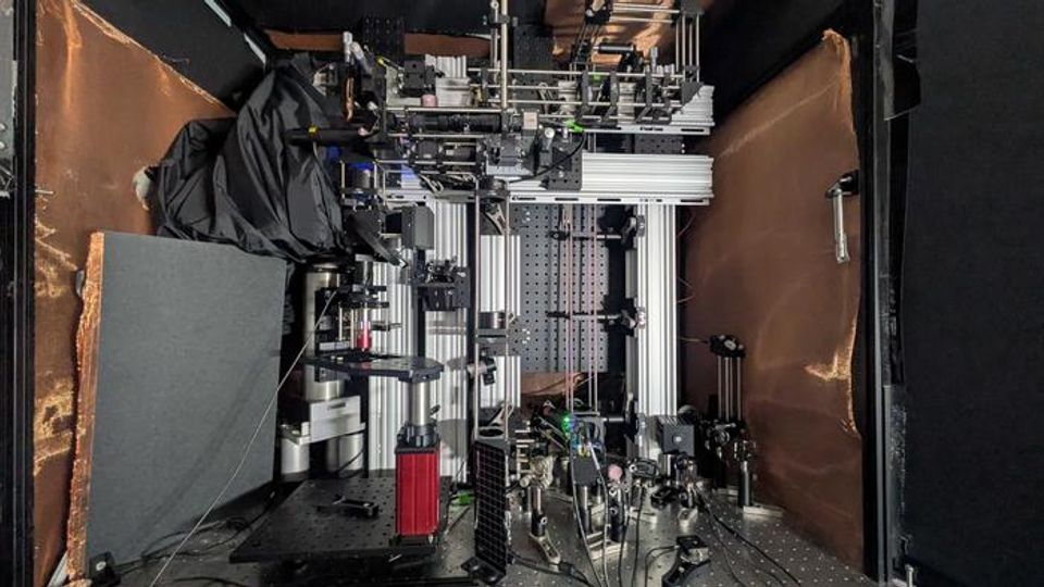
The system builds on three-photon microscopy but integrates a method for detecting sound waves generated by molecular interactions, enabling imaging through more than 1 mm of dense brain tissue. This is significantly deeper than the range achievable with traditional optical techniques.
Photoacoustic imaging enables deep, label-free microscopy
The research, published in Light: Science and Applications, details how the team imaged NAD(P)H – an intrinsic marker of cellular metabolism – through a 1.1 mm thick human-derived cerebral organoid and a 0.7 mm mouse brain slice.
Conventional methods excite NAD(P)H using ultraviolet light. In contrast, the MIT system uses femtosecond pulses of light at a wavelength three times longer than the molecule’s peak absorption. This “three-photon” excitation minimizes light scattering and penetrates deeper into tissues. The interaction produces localized heating within cells, which in turn creates ultrasonic pressure waves.
Photoacoustic imaging
A biomedical imaging technique that converts absorbed light into sound waves.
Third-harmonic generation imaging
An imaging technique that captures signals from the interface between cellular structures.
Instead of relying on emitted light, which can degrade rapidly at depth, the system detects these pressure waves using a highly sensitive ultrasound microphone. These acoustic signals are then reconstructed into high-resolution images. The researchers refer to this approach as “three-photon photoacoustic imaging.”
“The major advance here is to enable us to image deeper at single-cell resolution.”
Dr. Mriganka Sur.
The system also supports simultaneous third-harmonic generation imaging, allowing visualization of cellular structures. The combined outputs provide spatial and metabolic information at single-cell resolution.
"We integrated all these cutting-edge techniques into one process to establish this ‘Multiphoton-In and Acoustic-Out’ platform.”
Dr. Tatsuya Osaki.
Moving toward live-animal and clinical applications
The research was led by a multidisciplinary team including neuroscientists, mechanical engineers and biophysicists at MIT’s Picower Institute for Learning and Memory. The project was co-led by postdoctoral researchers and research scientists from laboratories directed by Professors Peter So and Mriganka Sur.
While current experiments used in vitro and ex vivo samples, the team plans to extend the system to in vivo imaging. A technical challenge in live tissues is aligning both the light source and the microphone on the same side of the sample, which differs from the setup used in static samples. However, based on current depth performance, the researchers believe that imaging up to 2 mm in live brains is feasible.
Beyond neuroscience research, the label-free system may have potential clinical uses. NAD(P)H levels are known to vary in neurological conditions such as Alzheimer’s disease, Rett syndrome and epilepsy. Because the method avoids external labels, it may be adaptable for intraoperative imaging during brain surgery.
The authors also suggest that other molecular targets, including genetically encoded indicators like GCaMP, could be detected using the same approach, extending the utility of the system to additional signaling pathways.
Reference: Osaki T, Lee WD, Zhang X, et al. Multi-photon, label-free photoacoustic and optical imaging of NADH in brain cells. Light: Science & Applications. 2025;14(1):264. doi: 10.1038/s41377-025-01895-x
This article has been republished from the following materials. Note: material may have been edited for length and content. For further information, please contact the cited source. Our press release publishing policy can be accessed here.
This content includes text that has been generated with the assistance of AI. Technology Networks' AI policy can be found here.


